What are the clinical symptoms of a Thelazia callipaeda infection?
Patients suffering from Thelazia callipaeda infection typically report persistent ocular discomfort characterized by the sensation of a foreign object in the eye. In the documented case of a 71-year-old crop farmer in South Korea, the symptoms lasted for approximately two months. The patient presented with significant redness and blurred vision, which are common indicators of ocular irritation caused by the presence of moving parasites.
Clinical examination by ophthalmologists revealed several physical abnormalities in the affected eyes. The conjunctiva, which is the membrane covering the eye, showed enlarged blood vessels, contributing to the visible redness. Furthermore, the cornea—the transparent outer layer of the eye—showed signs of erosion. This tissue damage is often a direct consequence of the physical movement and presence of the helminths within the ocular environment.
Physical manifestations of ocular parasitism
The presence of mobile, whitish roundworms within the eye causes mechanical irritation to the delicate tissues. This irritation can lead to inflammation of the conjunctiva and potentially more severe damage to the corneal cells. Because the worms are mobile, the sensation of something 'crawling' or being 'stuck' is a hallmark of the condition, making it difficult for patients to ignore the infection for extended periods.
How does the life cycle of Thelazia callipaeda function?
The life cycle of Thelazia callipaeda is complex and relies heavily on an insect vector to move between hosts. These nematodes primarily infest the eyes of domestic animals, such as cats and dogs, but can inadvertently infect humans. The process begins when flies feed on the tears and secretions of an infested animal, inadvertently consuming the parasite's larvae.
Once ingested by the fly, the larvae undergo a specific developmental process. According to the case report, the larvae molt twice within the insect, transforming into third-stage larvae. These larvae then migrate to the fly's mouthparts. When the fly next lands on a suitable host—ideally a watery eyeball—the larvae disembark and enter the host's eye. Once inside, they undergo one final molt to become adults, at which point they settle into the eye to live and mate.
The role of the insect vector
Flies act as both an incubator and a transport mechanism for the larvae. Without the intermediary insect stage, the parasite would be unable to navigate from one host to another. This biological dependency makes the management of fly populations in areas where these worms are endemic a significant factor in controlling the spread of the infection.
What is the biological distinction between male and female worms?
The Thelazia callipaeda worms exhibit distinct morphological differences between sexes, which are critical for identification under microscopic examination. Male worms are characterized by the presence of copulatory spicules, which are needle-like structures used to facilitate mating. In the South Korean case study, these structures allowed clinicians to definitively identify the sex of the extracted nematodes.
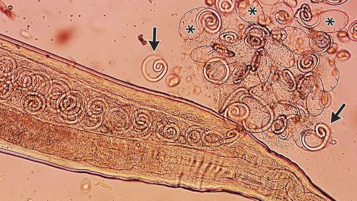
Female worms are notably different due to their ovoviviparous nature. Unlike many other parasitic species, female T. callipaeda develop embryos within eggs inside their own bodies. This means they do not lay eggs in the traditional sense but instead release large broods of densely coiled, first-stage larvae. In the medical case, some female worms were observed to be 'plump' with embryonated eggs, and larvae were seen bursting from them immediately after extraction.
| Feature | Male Worms | Female Worms |
|---|---|---|
| Key Identification Mark | Copulatory spicules | Embryonated eggs/Larvae |
| Reproductive Method | Mating via spicules | Ovoviviparous (larvae release) |
| Physical Appearance | Ventrally curved posterior | Plump with internal brood |
How is the infection treated and removed?
Treatment for a Thelazia callipaeda infection often involves direct mechanical extraction rather than systemic medication. In the case of the 71-year-old patient, ophthalmologists utilized a topical anesthetic to numb the eyes before beginning the procedure. This allowed the doctors to use fine forceps to pluck the 11 adult worms from the ocular tissue one by one.
The surgical removal of the adult worms is often sufficient to resolve the infection. Because the primary goal is to remove the breeding adults, the patient in the case study did not require antiparasitic medication, even though some larvae may have been left behind in the eye. The effectiveness of this approach was confirmed during a follow-up appointment two months later, where doctors noted that all ocular symptoms had resolved and no further worms were present.
Post-extraction recovery and monitoring
Following the removal of the parasites, the ocular tissues require time to heal from the inflammation and corneal erosion caused by the worms. Regular follow-up appointments are essential to ensure that no secondary infections occur and that the corneal integrity is restored. In the reported case, the patient showed a full recovery within a two-month window.
Frequently asked questions
What are the most common hosts for Thelazia callipaeda?
The primary hosts for these nematodes are domestic animals, specifically cats and dogs. These animals provide the necessary ocular secretions for the flies to feed upon, which facilitates the parasite's life cycle. Humans are considered accidental hosts when flies transfer the larvae from animals to human eyes.
Can eye worms be spread directly from person to person?
No, Thelazia callipaeda cannot be spread directly through human-to-human contact. The infection requires an intermediate insect vector, such as a fly, to transport the larvae from one host to another. The cycle depends on flies feeding on the secretions of an infested host.
Is the infection permanent if not treated?
If left untreated, the infection can lead to significant ocular damage, including corneal erosion and persistent inflammation. While the worms are mobile, their presence causes continuous mechanical irritation. Medical intervention is necessary to remove the adults and prevent the continuous release of larvae into the eye.
How do doctors identify the species under a microscope?
Doctors identify the species by examining the morphological characteristics of the worms. For T. callipaeda, this includes looking for specific structures like the copulatory spicules in males or the presence of densely coiled first-stage larvae within the uteri of the ovoviviparous females.
Do all patients require medication after worm removal?
Not necessarily. In many cases, such as the one reported in the New England Journal of Medicine, the physical removal of all adult worms is sufficient to end the infection cycle. This prevents further larvae from being released, allowing the eye to heal naturally without systemic antiparasitic drugs.
Key takeaways
- Thelazia callipaeda is a nematode parasite that inhabits the eyes of humans and animals.
- Infection is transmitted via flies that act as intermediate vectors for the larvae.
- Female worms are ovoviviparous, releasing live larvae rather than traditional eggs.
- Clinical symptoms include redness, blurred vision, and the sensation of foreign bodies.
- Mechanical extraction with forceps is a highly effective method of treatment.
Conclusion
The case of the 71-year-old farmer highlights the clinical realities of Thelazia callipaeda infection, a condition that, while rare in humans, presents significant ocular challenges. The parasite's ability to utilize insect vectors and its unique ovoviviparous reproductive strategy allow it to thrive in ocular environments. While the physical presence of the worms causes noticeable inflammation and corneal damage, prompt medical intervention through mechanical extraction can successfully resolve the infection. Understanding the life cycle and morphological characteristics of these nematodes is vital for accurate diagnosis and effective clinical management in affected regions.